Undertaking investigations will be a key part of understanding why you have spinal-related symptoms. For the majority of our patients, we will recommend an appropriate investigation after your initial consultation. However, we will also be happy to review any investigations you have previously had carried out prior to your consultation with us. Occasionally, we will need to repeat these or request complimentary further investigations as required.
A description of each of the investigations we use and the potential diagnosTic information they provide is described below:
1. MR Scan
Magnetic resonance (MR) imaging is almost always the preferred investigation for spine disorders. It provides excellent definition of the structures of the spine including the discs, the quality of the bone, the state of the nerves and the spaces within the spine in which they travel, as well as the joints and soft tissues around the spine.
MR images are generated by placing you in a very strong magnetic field. As a consequence of this, there are certain factors that preclude an MR scan being carried out, most notably the presence of metal inside your body whether this be in the form of a pacemaker or metallic foreign body. You will be asked to fill in a safety checklist before having an MR scan. MR scans will typically last anywhere from 20 to 45 minutes depending upon what sequences need to be carried out. You will be guided through the investigation by the MR radiographers. The other things you will notice about the scanner is how noisy it is and it is also a relatively confined space. If you have concerns about claustrophobia, please do mention this to your surgeon as scans can be done with sedation to make the experience as comfortable for you as possible.
2. CT Scan
A CT scan uses x-ray radiation to image the spine. It is particularly helpful in imaging the bony anatomy of the spine given how well this shows up on a CT scan. However, it is relatively poor at showing the nerves and the spinal cord within the spine, hence MR scanning is often preferred. However, if your surgeon does require information about the quality of bone or further resolution of bony structures such as facet joints, a CT scan can prove a very useful investigation.
A CT scan uses x-ray radiation but assuming that this will be a one-off investigation, the dose of radiation you receive will be very small. A CT scan is a lot quicker than an MR scan and also does involve the confined space of going into an MR scanner.
Occasionally, a CT scan will need to be done if there is contraindication to an MR scan. With the resolution of modern CT scanners this will very often provide a satisfactory alternative to an MR scan when required.
Very rarely, a dye can be injected into the spinal canal when having a CT scan to achieve better definition of the nerves. This is a CT myelogram but its use is very limited in current practice.
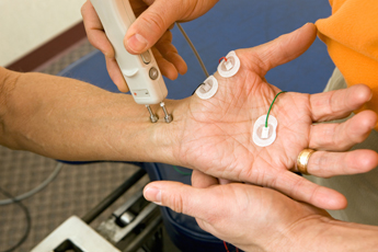
3. Nerve Conduction Studies and EMG (Electromyography)
This is a technique that is used to assess the functioning of the nerves of the peripheral nervous system, as well as the function of the muscles that they innervate. This investigation can be requested for a variety of reasons. In spinal disorders, it can help localise a level that may be symptomatic if your scan has shown spondylosis at multiple levels in the spine. It can also establish whether there is peripheral nerve compression contributing to your symptoms, such as carpal tunnel syndrome or ulnar nerve compression syndrome (cubital tunnel syndrome). It will on occasion also detect non-compressive problems of nerves such as peripheral neuropathies where there is an intrinsic abnormality of nerve function.
Nerve conduction studies and EMGs are carried out by highly trained specialist doctors who specialise in neurophysiology. The investigation is done by placing electrodes on the skin over nerves and stimulating them with small electrical currents. The speed of nerve conduction can then be calculated and this provides a lot of information about the state of nerve function. The EMG recordings are done by placing fine needles into the muscle and detecting how they respond to nerve stimulation.
4. Plain X-rays
Sometimes, plain x-rays can be helpful in investigating or monitoring a spinal condition. The most common situation in which x-rays are used is to establish whether there is any abnormal movement in the spine. These are referred to as dynamic x-rays and are taken with your spine in various positions. X-rays are also useful in determining the position of any implants that may have been used in previous spinal surgery